
A deep-learning model for thyroid cancer histopathology.
Built with a university hospital: a neural network that classifies thyroid cancer cells from histopathology images, tile by tile across a whole slide.
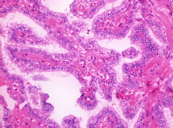
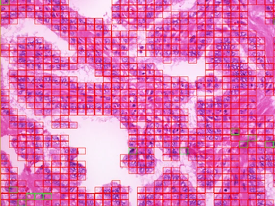
A sample run on one whole-slide image. Tile counts are that image’s output, not a measure of model accuracy.
We partnered with a university hospital that brought the clinical questions and the annotated histopathology data. We brought the machine-learning engineering. The goal was a model that could identify and classify thyroid cancer cells in tissue images.
A deep neural network, engineered in MATLAB, that divides each slide into a grid of tiles, classifies every tile as benign or malignant, and rolls the tiles up into a slide-level read, with the malignant regions annotated back onto the original image.
The model classifies whole-slide images and reports a slide-level impression with the malignant regions mapped. Measured performance is reported in the forthcoming paper rather than asserted here.
This is the depth we bring to any domain. If the same engineering can be trusted with cancer histopathology in a hospital setting, the systems we build for clinics, contractors, and logistics stand on serious ground.
The outcomes are being prepared for publication as part of an upcoming academic paper. We’ll link it here when it’s out.
Hospital partner cleared to be referenced and kept unnamed pending naming approval. This is research, not a cleared clinical diagnostic device; performance figures will publish with the paper.